Blocked Tear Ducts – Adults Dacryocystorhinostomy (DCR)
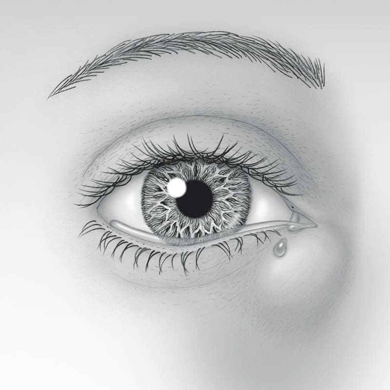
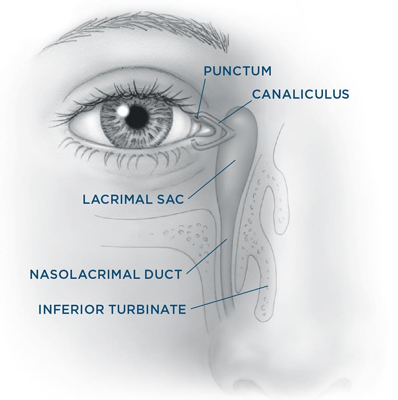
The tear drain starts with two small openings called puncta; one punctum is in the inner upper eyelid and the other is in the inner lower eyelid. You can see them with the naked eye if you look closely. Each of these openings leads into a small channel called the canaliculus which in turn empties into the lacrimal sac between the inside corner of your eye and nose. The lacrimal sac narrows into a tunnel called the nasolacrimal duct that passes through the bony structures surrounding your nose and then empties tears into your nasal cavity.

When you blink, your eyelids push tears evenly across the eyes to keep them moist and healthy. Blinking also pumps your old tears into the puncta and lacrimal sac where they travel through the nasolacrimal duct and drain into your nose.
The most common symptoms of blocked tear ducts include mucus buildup at the inside corner of the eye and/or along the lashes, excessive watering down the cheek (causing a need to wipe almost constantly throughout the day), and distorted vision. Depending on where along the passageway from the punctum to the nose the blockage occurs, you may also have redness, tenderness, and swelling between the inner corner of the eye and the side of the nose. An assessment by your oculofacial plastic surgeon can usually identify if a narrowing or blockage exists and if so, where along the passageway it is.

Treatments
Your surgeon may recommend a number of treatments based on your symptoms and exam findings. In some cases, it may be as simple as applying warm compresses and antibiotics but often, surgery is needed.
Narrowing of the punctum or canaliculus may respond to minor office procedures to reopen these passageways (snip punctoplasty or stenting procedures). A common location of blockage is the nasolacrimal duct. This causes tears to be trapped in the lacrimal sac and sometimes become stagnant and infected. If the nasolacrimal duct is narrowed (also known as “stenosis”) but still partially open, your surgeon may recommend placing temporary stents through your nasal passageways. If this is not effective or if the nasolacrimal duct becomes completely blocked, a dacrocystorhinostomy (DCR) is the gold standard surgery to correct this problem.
To perform a DCR, your surgeon will create a new drainage opening from the blocked sac directly into your nose to bypass the obstruction in your nasolacrimal duct. (In children, in whom a blocked nasolacrimal duct can often be opened with a probe, but adults are usually not able to be treated in this way.) A small incision is made either in the skin or inside the nose. A fine, soft silicone stent may temporarily be left in the new tear drain for a few weeks to keep the duct open while healing occurs.
A DCR is an outpatient procedure that may be done under twilight sedation or general anesthesia. You may have mild nose bleeding for a couple of days after the procedure. Most people recover within days.
A less common site of complete blockage is at the level of the canaliculus. In this situation, a prosthetic glass tube (often referred to as a Jones tube) may be placed to serve as a gutter to catch tears from the surface of the eye and shunt them into the nasal cavity. This procedure is also called a conjunctivodacryocystorhinostomy (CDCR). Unlike a DCR that requires virtually no maintenance after surgery, a glass tube will likely need some degree of cleaning, repositioning, or replacement of the tube.
Most patients experience improvement in their tearing and discharge once the appropriate procedure has been done to address the blockage in their tear drainage system.
Risks and Complications
Bleeding and infection are potential risks, as with any surgical procedure. Minor bruising and swelling between the eye and nose should be expected and will likely go away in one to two weeks. Passageways may narrow again or scar tissue may grow around the new opening made in a DCR or CDCR. Further surgery may be required.
Patients requiring a continuous positive airway pressure (CPAP) device for sleep apnea should notify their surgeon because they may experience air on the eye after successful tear drain surgery. Your surgeon cannot control all the variables that may impact your final result. There is always a chance that tear drainage surgery is not successful and additional treatment is needed or tearing may persist.